A primary cell culture model of rabbit uroepithelium
Isolation of Epithelial Cells from Rabbit Bladders
1. Animal experiments were performed in accordance with the Animal Use and Care Committee.
2. Urinary bladders were obtained from New Zealand White rabbits (3–4 kg).
3. Usually, two rabbits were used per culture.
4. Each rabbit was euthanized with 250 mg of pentobarbital, the bladder was exposed, the ends of the bladder were clamped with hemostats, an incision was made lengthwise along the bladder and the opened bladder was surgically excised and washed in Krebs solution (110 mM NaCl, 5.8 mM KCl, 25 mM NaHCO3, 1.2 mM KH2PO4, 2.0 mM CaCl2, 1.2 mM MgSO4, 11.1 mM glucose, pH 7.4).
5. The bladder was trimmed of excess fat and stretched on a rack, mucosal side down, in the same solution at 37 °C.
6. The smooth muscle layers were carefully removed by disscaling with a scalpel and forceps and the stripped mucosa was transferred to a 10 × 10-cm square dish containing an 8 × 8-cm plastic rack with 10 sharp metal pins along each edge.
7. The tissue was stretched mucosal side up across the metal pins and then incubated overnight at 4 °C in sterile minimal essential medium containing 1% (v/v) penicillin/streptomycin/fungizone, 2.5 mg/ml dispase, and 20 mM HEPES , pH 7.4, to weaken the association between the epithelium and the connective tissue and to facilitate manual separation of the mucosa.
8. Following treatment, the stripped mucosa was transferred to a sterile tissue culture hood, the minimal essential medium/dispase solution was aspirated, and the epithelial cells were scraped from the underlying connective tissue using two flexible cell scrapers.
9. The scraped cells were transferred to a tissue culture dish, resuspended in 20 ml of 0.25% (w/v) trypsin-1 mM EDTA, and incubated 15–30 min at 37 °C, resuspending once during the incubation.
9. The scraped cells were transferred to a tissue culture dish, resuspended in 20 ml of 0.25% (w/v) trypsin-1 mM EDTA, and incubated 15–30 min at 37 °C, resuspending once during the incubation.
10. After trypsinization, the single cell suspension was brought up to 50 ml with minimal essential medium containing 1% (v/v) penicillin/streptomycin/fungizone, 20 mM HEPES, pH 7.4, and 5% (v/v) fetal bovine Serum in a sterile conical tube and spun down in an IEC Centra CL2 Centrifuge at 1,000 rpm for 5 min to pellet the cells and remove the trypsin.
11. The supernatant was aspirated carefully and the cells were resuspended in 50 ml of the same minimal essential medium/penicillin/streptomycin/fungizone/fetal bovine serum solution and washed an additional two times.
The cells were then washed in 50 ml of defined keratinocyte medium and then resuspended in the appropriate volume of defined keratinocyte medium to yield a final concentration of 700,000–800,000 cells/ml, as determined by cell counting in a hemocytometer chamber.
13. This cell density was determined to obtain highly differentiated cultures as plating at a higher (≥900,000 cells/ml) or lower density (<500,000 cells/ml) resulted in poorly developed cultures.
14. Two bladders yielded enough cells to plate approximately 50–60 12-mm Transwell filters.
Plating and Cell Culture
1. Cells were plated on either 12-mm Transwells or 12-mm Snapwells coated with collagen.
2. The collagen solution was prepared by mixing 10 mg of type IV collagen, 200 μl of glacial acetic acid, and 100 ml of H2O and incubating overnight at 4 °C without stirring.
3. The collagen solution was sterile filtered and stored at 4 °C.
Prior to use, the collagen solution was diluted 1:9 in 10 mM Na2CO3-HCl, pH 9.0, and 500 μl of the resultant solution was added to each filter and incubated for 60 min at room temperature to allow the collagen to bind to The filters.
5. Prior to plating, the collagen solution was aspirated and 0.5 ml of cell suspension was added to the apical chamber and 2 ml of keratinocyte medium was added to the basal chamber.
6. For Snapwells, 0.5 ml of the cell suspension was added to the apical chamber and 4 ml of keratinocyte medium was added to the basal chamber.
7. The third day after plating, the apical medium was aspirated and replaced with 1 ml of keratinocyte medium for the Transwells and 0.5 ml of medium for the Snapwells.
8. The basolateral medium was exchanged for 1.5 or 4 ml of defined keratinocyte medium per Transwell or Snapwell, respectively.
9. The cells were fed in this manner every 2–3 days.
10. Cells whose TER reached levels of approximately 200 Ω cm2 or higher (between 3 and 6 days after plating) were switched to keratinocyte medium containing 1 mM calcium chloride in order to achieve high TER (8000 Ω cm2 or greater).
11. Adding 1 mMcalcium chloride before day 3 resulted in poorly developed cultures, while adding calcium chloride after day 6 resulted in cultures with suboptimal TER (<8000 Ω cm2).
12. Successful cultures were attained approximately 85% of the time.
Product Description:
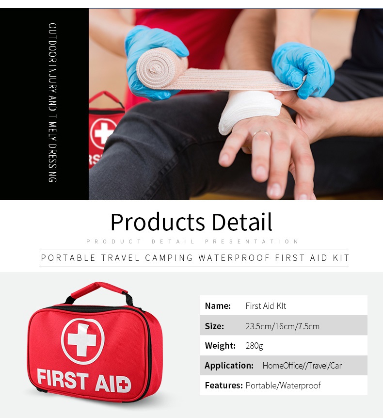
✓ COMPREHENSIVE. It has the perfect selection of emergency and survival supplies. You'll have peace of mind knowing that your kit is packed with everything you need in times of crisis.
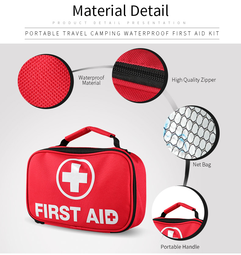
✓ DURABLE BAG. This kit is designed to be tough, it is crafted with ripstop fabric and strong zipper, which provide that extra level of durability and protection.
✓ YOU CAN TAKE IT ANYWHERE. This kit is made to get you through a wilderness trip, a drive across the country, or just around your own home for daily misshapes.
✓ COMPACT. The internal layout is organized whilst the pack is durable and fairly lightweight. Ideal for Car, Home, Office, Camping, Travel, Sports, and more. Includes a mini first aid kit.


Dongguan Maimeng Culture and Creative Co. Ltd , https://www.factorydirectmask.com