At Azure Biosystems, we strongly believe in the power of fluorescent multiplexes in Western blots, which has led to the rapid development of our Azure C-Series imagers. But we realize that taking the standard HRP/chemiluminescence method is a bit daunting, so we have proposed six considerations to make the conversion of chemiluminescence to fluorescent westernblot as simple as possible.

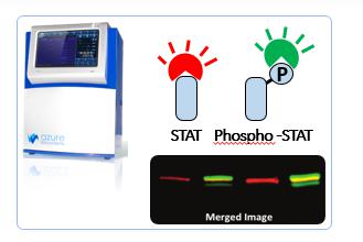
We also covered the relevant content in some of our previous applications, such as phosphoric acid / total protein detection, etc.
1. Increased concentration - The concentration of primary and secondary antibodies required may increase compared to chemiluminescent Western blots, depending on the imager system you are using. In more modern or laser imagers, this effect may be less noticeable.
2. First test the monochrome experiment - it is very meaningful to test your antibody separately. This way, you can determine the optimal concentration, taking into account background or non-specific factors in a simple environment, rather than analyzing 3 different primary and secondary antibodies at once.
3. Using Adsorbent Secondary Antibodies - Although it sounds simple, many people don't consider that they now add multiple antibodies and multiple species to a single blot. If you use multiple fluorescences in other areas, you will already be aware of the cross-adsorption of secondary antibodies and the importance of reducing cross-reactivity between species. But in the West, many people don't think about it, it's worth checking to make sure they meet the requirements.
4. Extended Spectrum - Three-color western blots are exciting, and that's five colors. Using the NIR function, you can greatly increase the distinction between different peaks. Obviously, this may require some work for antibody optimization, and once for the detection of 5 proteins can save time and cost.
5. Check your blotting membrane - Although more and more fluorescent safety membranes are under development, some membranes emit autofluorescence under UV light source exposure to produce high background signals. We also recommend switching from the NC membrane to the PVDF membrane for increased sensitivity, as described previously.
6. Choose the right channel for your protein - all detection channels are not created equal. For standard fluorescence, use the blue channel to detect your highest abundance protein, the green channel to detect medium abundance proteins and the red channel to detect low abundance proteins. If NIR is used, excellent sensitivity and low background are also ideal for low abundance proteins.
If you follow these tips and read some of our application guides, the transition from single to multi-channel should be very smooth, and we are always ready to provide you with technical support if you encounter any difficulties.
Liquid-based Cytology Screening
TCT
Hangzhou DIAN Biotechnology Co., Ltd. , https://www.dianbiotech.com